

3D cell culture for studying cell behavior
Precisely fabricated 3D cell scaffolds provide defined geometries and physical cues for studying cell adhesion, migration, differentiation, and responses to their surroundings. Quantum X bio enables researchers to create these cell microenvironments with sub-cellular resolution, 3D design freedom, and controlled process conditions.
Engineering 3D microenvironments for cell studies
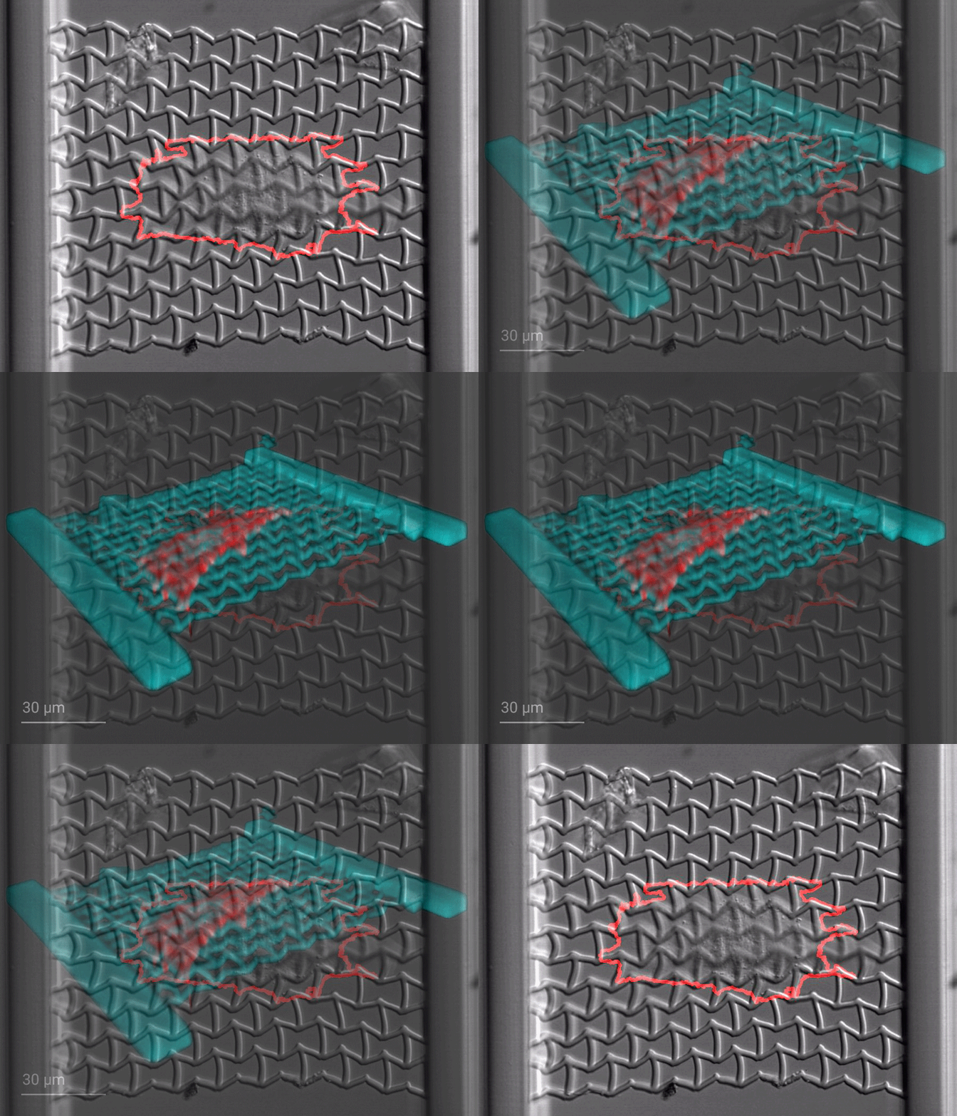
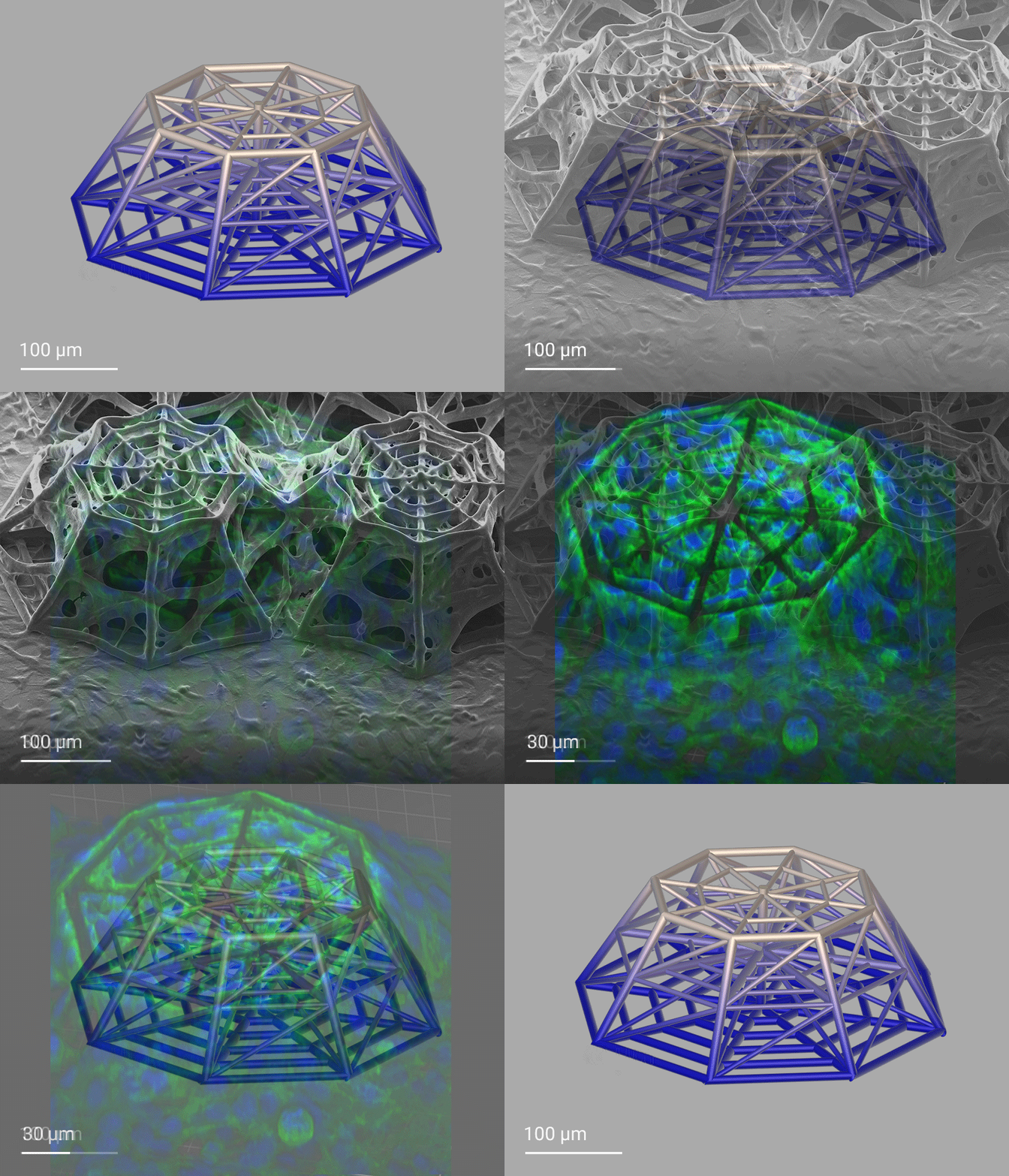
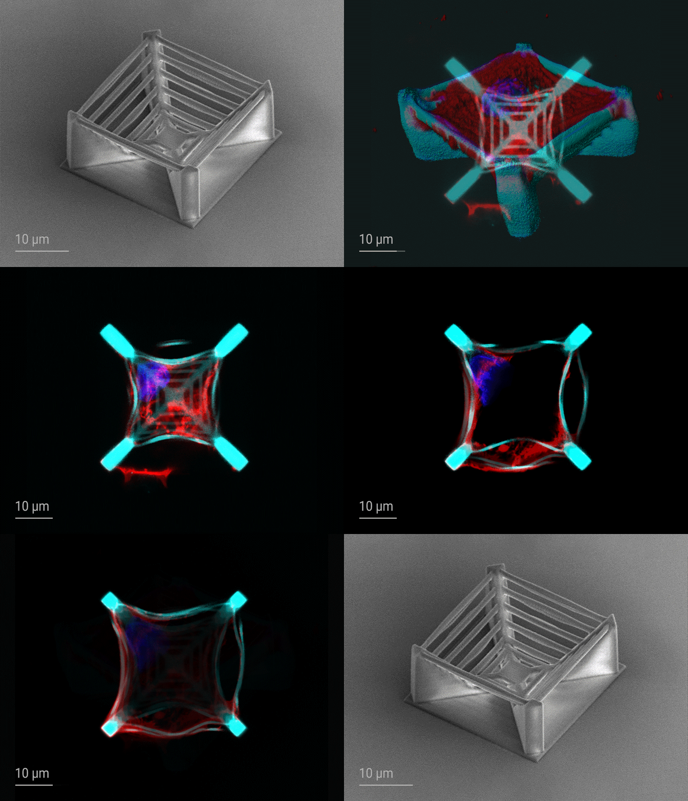
3D cell culture for reproducible in vitro cell studies
Cells behave differently in conventional 2D cultures than in living tissue, where cell adhesion, force sensing, and cell migration are shaped by complex three-dimensional surroundings. In 3D cell culture, engineered cell scaffolds allow scientists to systematically vary pore size, spacing, and confinement to study how defined geometric cues affect cell behavior. These cell microenvironments support studies in mechanobiology, stem cell biology, and neural or vascular models for investigating cell differentiation and disease progression.
Engineering cell-scale microenvironments with geometric precision
3D cell culture relies on engineered microarchitectures such as porous cell scaffolds, microcages, capillary-like networks, and microfluidic perfusion channels that influence cell adhesion, migration, proliferation, differentiation and force generation in defined environments. Precise manufacturing technology is critical because scaffold performance depends on geometric precision at the cell scale, including tailored surface topographies, free-form features, and reproducible scaffold networks. Nanoscribe's Quantum X systems combine 3D design freedom with control over cell-scale features and pore dimensions, helping researchers fabricate highly tailored 3D cell culture scaffolds for studies under defined conditions.
Why Nanoscribe for 3D cell culture
Nanoscribe’s Quantum X bio enables precise 3D cell scaffold fabrication under controlled bioprinting conditions.
-
Fast, high-precision 3D scaffold fabrication: With 3D printing by Two-Photon Grayscale Lithography (2GL®), Quantum X bio enables rapid fabrication of cell scaffolds and related microstructures ranging from subcellular features to millimeter-sized structures.
-
Cell-scale geometry control: Dynamic voxel size control and submicron-resolution printing enable finely resolved, reproducible scaffold architectures and controlled pore geometries in all three dimensions, while maintaining high shape accuracy, and high-resolution surface features.
-
Controlled bioprinting environment: A bioprinting chamber controls temperature and humidity and provides HEPA-filtered airflow and a connection for premixed air/CO2. Heaters in the Bioprinting Set control cell culture dish temperature.
-
Open material system: Third-party and custom materials can be developed, tested, and deployed quickly for application-specific scaffold fabrication and functional resin development.
-
Commercial material options: The platform supports biocompatible Nanoscribe photoresins, including stiff and flexible materials, and hydrogels from Advanced BioMatrix and BIO INX.
-
Flexible substrate support: The platform supports printing on microscope slides, cell dishes, and microfluidic chips for in-chip structures, tissue models, and organ-on-a-chip setups.
Your questions answered: 3D cell culture
Why are 3D cell scaffolds used in cell culture?
Cells respond strongly to the physical and biochemical properties of their surroundings. In conventional 2D cultures, cells grow on flat surfaces that lack the complex three-dimensional environments found in living tissues. Engineered 3D scaffolds introduce defined geometries and physical cues, enabling researchers to study cell adhesion, migration, differentiation, and mechanobiological responses in environments under controlled conditions. Nanoscribe's 3D printing by Two-Photon Grayscale Lithography (2GL®) enables cell-scale scaffold features and controlled pore geometries for such studies.
Why is fabrication precision important for 3D cell culture scaffolds?
Cells respond sensitively to their physical environment, making scaffold reproducibility and geometric accuracy critical. Small variations in pore size and spacing, surface topography, confinement or structural dimensions can influence cell behavior and experimental outcomes. High-precision scaffold fabrication helps create consistent cell microenvironments, enabling more reliable and comparable in vitro studies.
What materials are used for 3D cell culture scaffolds?
3D cell culture scaffolds can be fabricated from a wide range of materials, including biocompatible photoresins, hydrogels, and custom biomaterial formulations. For example, researchers printed retinal cell scaffolds from gelatin, and a gelatin/HA blend. Material selection depends on the application and desired properties, such as stiffness, degradability, transparency, cell adhesion, or biofunctionalization. Nanoscribe's Quantum X bio supports both commercial and custom materials, enabling researchers to tailor scaffold properties to specific cell types and tissue models. Varying these properties within a defined scaffold design helps researchers investigate how material properties influence cell behavior.
Read the related publication here (register for free):
Development of High-Resolution Three-Dimensional-Printed Extracellular Matrix Scaffolds and Their Compatibility with Pluripotent Stem Cells and Early Retinal Cells
How can 3D cell culture scaffolds be integrated with microfluidic devices?
Many researchers integrate 3D scaffolds into microfluidic chips to create controlled culture environments with defined fluid flow, nutrient transport, and chemical gradients. These systems support applications such as vascular models, organ-on-a-chip devices, and barrier tissues, providing more physiologically relevant conditions than static cultures. Nanoscribe’s Quantum X bio enables scaffold fabrication directly within microfluidic devices and other cell-culture platforms, supporting highly customized experimental designs.
Get your project started with our products
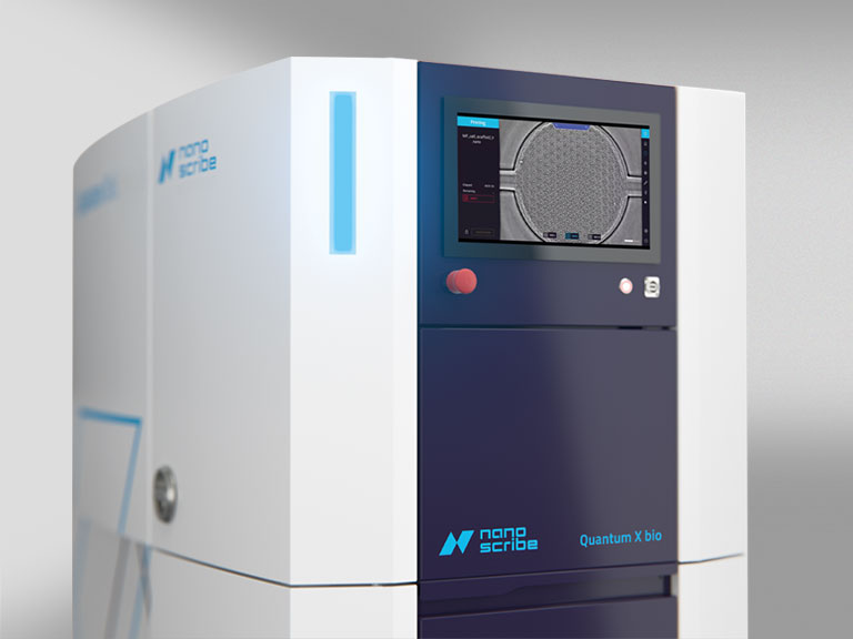
Quantum X bio
3D printing for life sciences and microfluidics
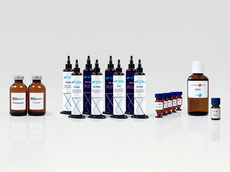
High-res 3D biofabrication materials
Materials for high-resolution 3D biofabrication in life sciences
Discover the potential of 3D cell culture scaffolds
Get inspired by these scientific highlights showcasing cell scaffolds created with Nanoscribe’s high-resolution 3D printing technology. For even more insights, explore over 2,500 peer-reviewed scientific publications in our premium resources section – simply log in or register for free.
Two‐Photon Laser Printing to Mechanically Stimulate Multicellular Systems in 3D
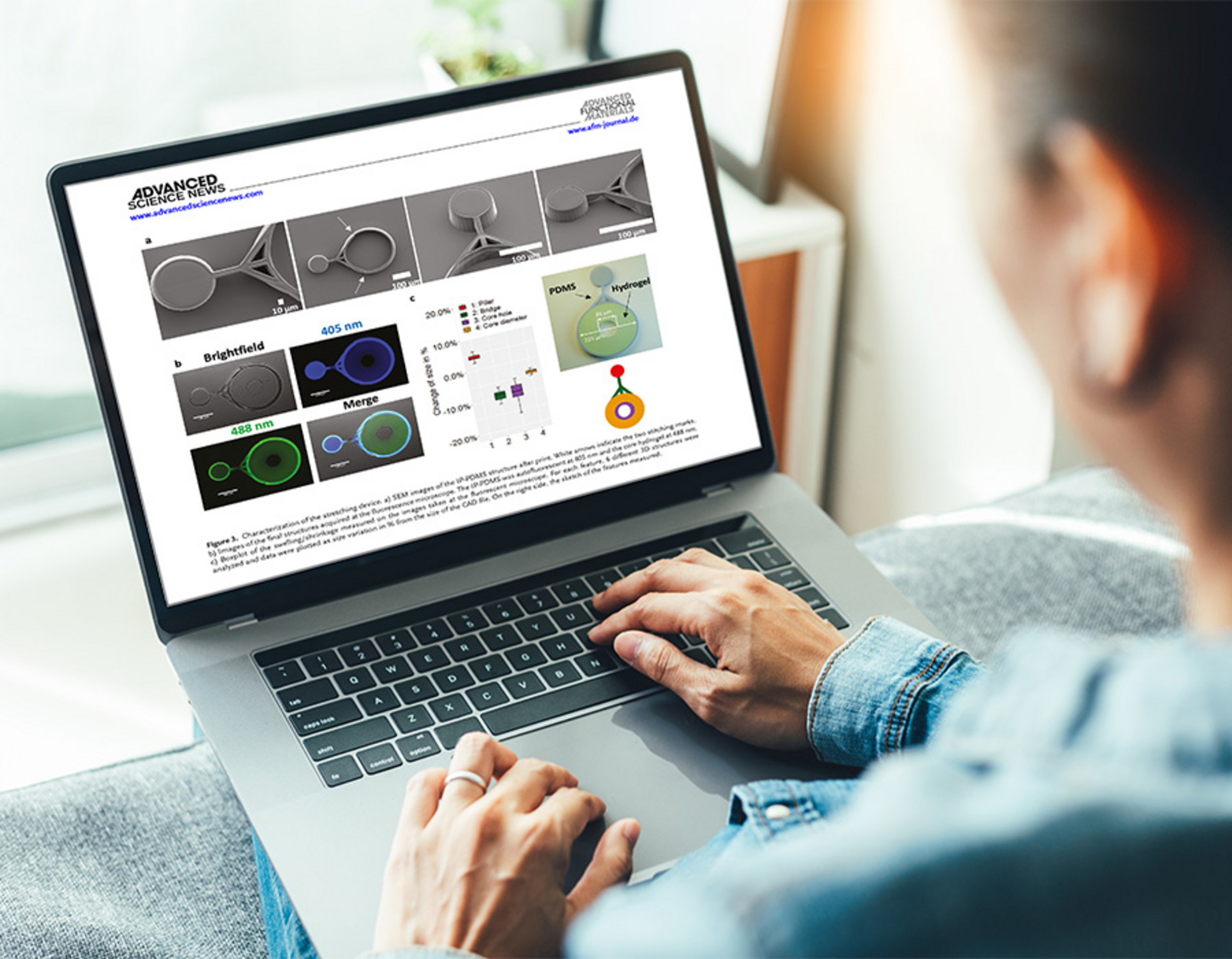
F. Colombo, M. Taale, F. Taheri, M. Villiou, T. Debatin, G. Dulatahu, P. Kollenz, M. Schmidt, C. Schlagheck, J. Wittbrodt, C. Selhuber‐Unkel
Universität Heidelberg, Heidelberg International Biosciences Graduate School HBIGS and HeiKa Graduate School on “Functional Materials”
Advanced Functional Materials, Volume 34, Issue 20 (2024)
Bio-Metamaterials for Mechano-Regulation of Mesenchymal Stem Cells
N. Munding, M. Fladung, Y. Chen, M. Hippler, A. D. Ho, M. Wegener, M. Bastmeyer, M. Tanaka
Heidelberg University, Karlsruhe Institute of Technology (KIT), Kyoto University
Advanced Functional Materials, Volume 34, Issue 19 (2023)
Evaluation of Proton-Induced DNA Damage in 3D-Engineered...
Q. Akolawala, M. Rovituso, H. H. Versteeg, A. M. R. Rondon, A. Accardo
Delft University of Technology, Holland Proton Therapy Center (HollandPTC), Leiden University Medical Center
ACS Applied Materials Interfaces 14 (18): 20778–20789 (2022)
Development of 3D culture scaffolds for directional neuronal growth using...
L. Agrawal, M. Saidani, L. Guillaud, M. Terenzio
Okinawa Institute of Science and Technology Graduate University
Materials Science and Engineering, Volume 131 (2021)